Kids Corner
Welcome to the Virtual Gabrieli Lab!
At the Gabrieli Laboratory, we study how the brain works. This page will help you learn more about YOUR brain, how it works, and what is like to participate in brain research.
How the Brain Works
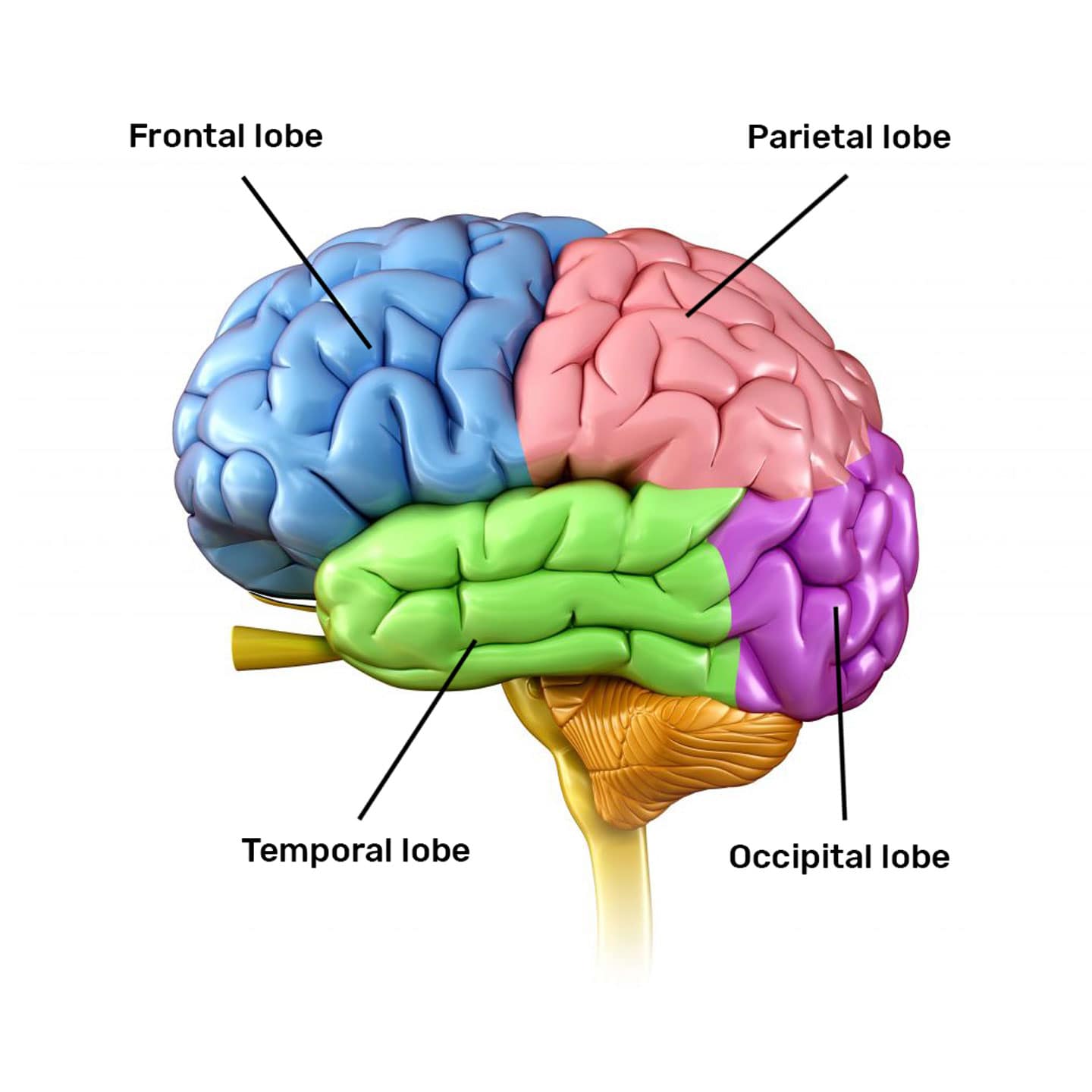
You know you have a brain, but do you know how it is structured and how it works? The brain is separated into four main regions – lobes – that each handle different mental tasks. The four lobes are the frontal, parietal, temporal, and occipital lobes.
- The frontal lobe, located in the front of the brain (the area behind your forehead) controls thinking, planning, problem-solving, short-term memory and movement.
- The parietal lobe interprets sensory information, such as taste, temperature and touch.
- The occipital lobe processes images from your eyes and links that information with images stored in memory.
- The temporal lobe processes information from your senses of smell, taste and sound. It also plays a role in memory storage.
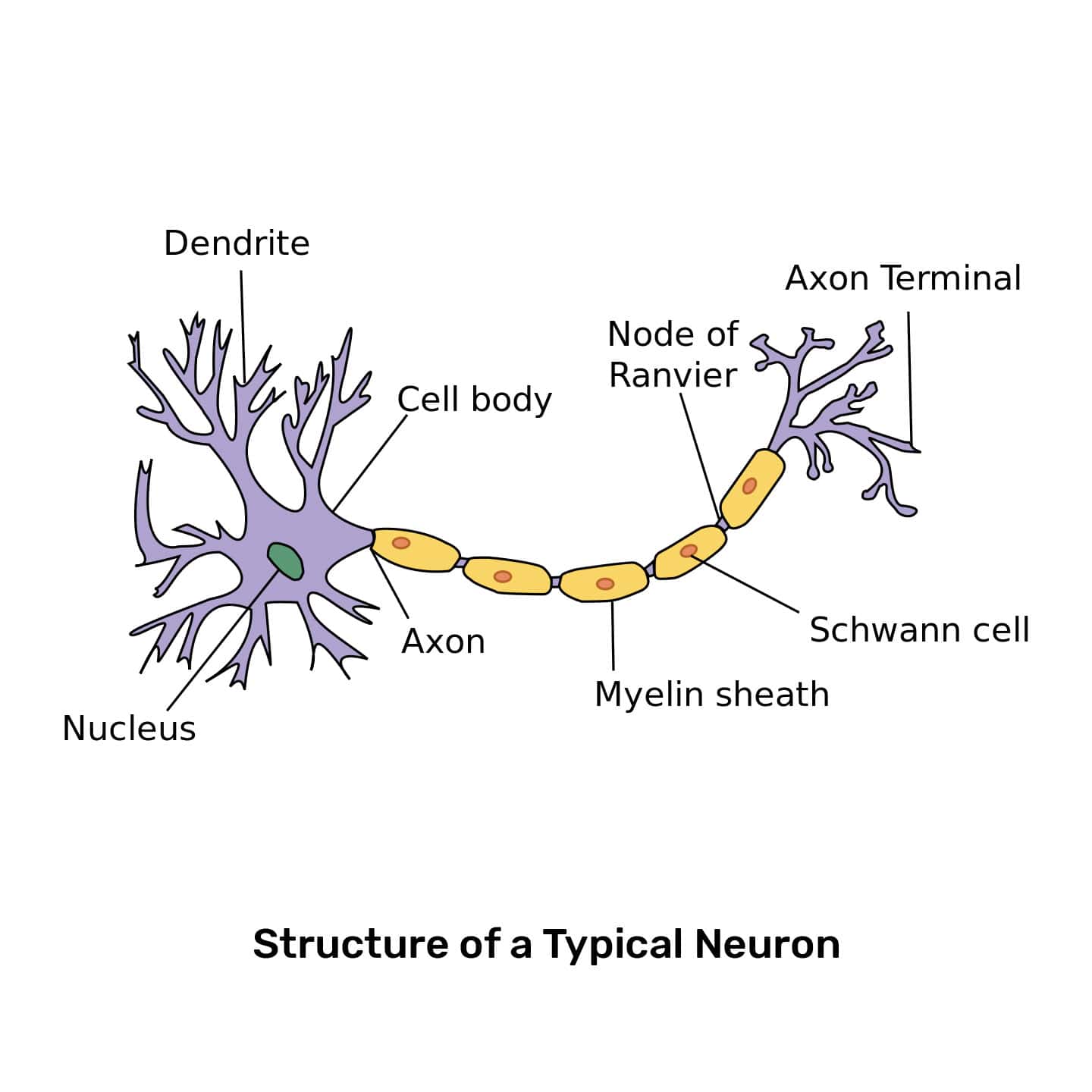
These lobes all communicate with each other in order to allow you to think, move, and feel. But how do they communicate? The brain talks with itself and with the rest of the body via cells called neurons. There are tons of these in your body; the human brain alone has approximately 86 billion neurons in it! The human Neurons use electrochemical signals to convey messages with other neurons using chemicals called neurotransmitters. When neurotransmitters are released by a neuron, the dendrites – branch-like receptors of neurotransmitters – of nearby neurons detect these signals.
When the nearby neuron detects enough of a neurotransmitter, it sends an electrical signal called an action potential down the axon (the long part of the neuron in the diagram). This is the electro aspect of electrochemical. The signal reaches the axon terminals and causes them to release neurotransmitters into an area called the synapse (the gap between two neurons). This is the chemical aspect of electrochemical. Synapses are tiny spaces; they are, on average, 20-40 nanometers (nm) wide. For perspective, the thickness of a piece of paper is around 100,000nm! A single neuron has thousands of synapses between itself and nearby neurons; a signal from one neuron can be picked up by many nearby neurons. There are always tons of neurons rapidly communicating with each other in your brain!


A Day with EEG
Electroencephalography (EEG) is a recording of the electrical activity in your brain produces. To be able to record this activity in your brain, we need to place a cap on your head. The cap is soft and stretchy and feels like a swim cap. This cap also has small holes in it, where we will place some gel and attach electrodes to your head. These “brain cell listeners” can pick up the electrical activity of your brain. This procedure is easy and completely safe, but the gel will make your hair messy.
What happens during an EEG session?
If you participate in an EEG study with us at Massachusetts Institute of Technology, you will come to the McGovern Institute for Brain Research.
- A researcher from the Gabrieli Lab will meet you and take you to our EEG lab, where you will learn how to play the computer games we have designed for you. You can ask as many questions as you like while you learn these games and set up for the EEG.
- After you have learned all of the computer games, one of our researchers will set up the EEG cap on your head. You might feel a slight tickle on your scalp while we place the electrodes and make sure the cap is secure. After the cap is in place, we are ready to start the experiment!
- Our EEG experiments usually last for 30 – 60 minutes. You will be playing computer games and watching movies while we record the electrical activity in your brain. After the experiment is over, you will receive Amazon gift cards for the time you spent with us at MIT!
Preparing for your EEG session at MIT
Please come with clean, dry hair to your EEG session. If you usually wear your hair in braids, please take them out first; this helps the cap fit better. Try to get a good night’s sleep the night before your EEG session and come ready to learn!
A Day with fMRI
MRI stands for Magnetic Resonance Imaging. Our MRI scanner is a big magnet that uses magnetic and radio waves to take pictures of your brain. Researchers at MIT take three types of brain pictures using the MRI scanner:
- Structural Images: These images show us the structure of your brain. At the end of your visit, you can take home these pictures of your brain on a CD to show your family what your brain looks like!
- Functional Images: fMRI stands for “functional” Magnetic Resonance Imaging, and means that we can take pictures of your brain while it is working on different tasks. During your scan, we will ask you to play computer games that we have designed so that we take fMRI pictures of your brain. These pictures show us which parts of your brain you are using while you play each game.
- Diffusion Tensor Imaging: A DTI scan is a special kind of scan that allows us to see the natural flow of water in your brain. These images help us understand how the white matter in your brain forms “highways” to connect different parts of your brain.
What happens during an fMRI session?
If you participate in an fMRI study with us at MIT, you will come to the McGovern Institute for Brain Research.
- A researcher from the Gabrieli Lab will meet you and take you to our fMRI scanning center, where you will have a chance to practice in our mock scanner and learn how to play the computer games we have designed for you.
- After you have learned all of the computer games, one of our researchers will take you into the real scanning room to get set up. The tunnel that you see is called the bore, and you will be lying inside the tunnel on a soft mattress while we take your brain pictures!
- A researcher will help you get onto the scanner bed, and we will give you some soft earphones so you can hear the sounds of the movies you will be watching! You will also be able to talk to and hear the research team during your scan using these headphones.
- During your scan, we will ask you to lie as still as possible so that we can take clear pictures of your brain. If you move, the pictures come out blurry! You will be watching movies and playing computer games throughout your time in the scanner.
Preparing for your fMRI session at MIT
Please come to your fMRI session well rested! It is important to get a good night’s sleep and eat a good breakfast before you come in so that we can take good pictures of your brain’s activity while you are playing games in the scanner!
It is also important to wear soft, comfortable clothes without metal (including zippers, buttons, or snaps) when you come in for your scan. Since the fMRI scanner is a giant magnet, you cannot have any metal on your body when you enter the scanning room. We will double check that you don’t have metal on your body by using a metal detector before you start your scan.
